Home » Without Label » Knee Muscle Anatomy Mri : Mri anatomy of knee Dr. Muhammad Bin Zulfiqar : Anatomical structures of the lower limb (hip, thigh, knee, leg, ankle and foot) and specific regions (compartment of the lower.
Knee Muscle Anatomy Mri : Mri anatomy of knee Dr. Muhammad Bin Zulfiqar : Anatomical structures of the lower limb (hip, thigh, knee, leg, ankle and foot) and specific regions (compartment of the lower.
Knee Muscle Anatomy Mri : Mri anatomy of knee Dr. Muhammad Bin Zulfiqar : Anatomical structures of the lower limb (hip, thigh, knee, leg, ankle and foot) and specific regions (compartment of the lower.. Superiorly, it extends to the level of the crossing of the biceps femoris tendon, and remains superficial to fcl in this location.10 This mri knee sagittal cross sectional anatomy tool is absolutely free to use. Magnetic resonance imaging is particularly well suited for the medical evaluation of the musculoskeletal (msk) system including the knee, shoulder, ankle, wrist and elbow. In one investigation, depicted only on the proton density weighted images. Articular muscle of the knee (articularis genu m.) normal mr imaging anatomy of the knee.
Three conventional mri planes that are utilized to evaluate the knee include sagittal (oblique), coronal, and transaxial planes. In one investigation, depicted only on the proton density weighted images. It is considered a vestigial muscle, and can be used as a tendon graft in reconstructive orthopedic surgery. Anatomy arthrogram anatomy basic shoulder mri. Thigh muscles are responsible for allowing normal gait and proper lower extremity function (1).
MRI KNEE JOINT ANATOMY from image.slidesharecdn.com This mri hip joint axial cross sectional anatomy tool is absolutely free to use. The medial thigh muscles are responsible for the adduction (movement of a body part toward the body's midline) of the leg. Anatomy of the ankle and foot in mri. Branches from the femoral, tibial, common peroneal, and obturator nerves; T2w axial fat sat 1. These motions of the knee allow the body to perform such important movements as walking, running, kicking, and jumping. While a detailed explanation of mri protocols and mr physics is beyond the scope of this text, fast spin echo (fse) mri is most commonly utilized for mri of the knee. Prescribe sagittal plane off axial images with line parallel to bony glenoid.
Radiology knee this app is a valuable tool for radiologists surgeons medical students doctors and nurses.
In conclusion, we describe the normal mri anatomy of the distal biceps femoris and the relationship of this muscle with the common peroneal nerve. This represents an accessory ossicle found in 10 30 of the normal population. The thigh has some of the body's largest muscles. Articular muscle of the knee (articularis genu m.) normal mr imaging anatomy of the knee. Two condylar joints between femur and tibia; The deepest layer consists of the popliteus muscle and its tendon passing. Thigh muscles are responsible for allowing normal gait and proper lower extremity function (1). It is considered a vestigial muscle, and can be used as a tendon graft in reconstructive orthopedic surgery. Abnormal anatomy with normal signal, i.e. Plantaris can have variable size, but in most cases is difficult to demonstrate on routine mri studies. Louis, usa and the rijnland hospital in leiderdorp, the netherlands. This mri hip joint axial cross sectional anatomy tool is absolutely free to use. Plantaris acts weakly to plantar flex the foot and flex the knee.
The muscles of the knee include the quadriceps, hamstrings, and the muscles of the calf. Anatomy basic knee mri checklist. Radiology knee this app is a valuable tool for radiologists surgeons medical students doctors and nurses. When interpreting the proton density images it. Superiorly, it extends to the level of the crossing of the biceps femoris tendon, and remains superficial to fcl in this location.10
Supplemental Materials for Normal MR Imaging Anatomy of ... from www.mri.theclinics.com Medical images from an mri allow medical professionals to distinguish body tissues, including the meniscus (shock absorbers in the knee), cartilage, tendons, and ligaments. Two condylar joints between femur and tibia; Rotation whilst in the flexed position to 10° actively and 60. Can also generate proton density images. Anatomical structures of the lower limb (hip, thigh, knee, leg, ankle and foot) and specific regions (compartment of the lower. Use the mouse scroll wheel to move the images up and down alternatively use the tiny arrows (>>) on both side of the image to move the images. Anatomy of the knee can be complicated and hard to understand. T2w axial fat sat 1.
The deepest layer consists of the popliteus muscle and its tendon passing.
They were created by volume rendering from a ct scan of the knee. In conclusion, we describe the normal mri anatomy of the distal biceps femoris and the relationship of this muscle with the common peroneal nerve. Three conventional mri planes that are utilized to evaluate the knee include sagittal (oblique), coronal, and transaxial planes. The thigh has some of the body's largest muscles. Articular muscle of the knee (articularis genu m.) normal mr imaging anatomy of the knee. The muscles of the knee include the quadriceps, hamstrings, and the muscles of the calf. There is a flat area of tendon originating from the knee. The deepest layer consists of the popliteus muscle and its tendon passing. Superiorly, it extends to the level of the crossing of the biceps femoris tendon, and remains superficial to fcl in this location.10 Two condylar joints between femur and tibia; Plantaris can have variable size, but in most cases is difficult to demonstrate on routine mri studies. This mri hip joint axial cross sectional anatomy tool is absolutely free to use. Anatomy basic knee mri checklist.
These motions of the knee allow the body to perform such important movements as walking, running, kicking, and jumping. Radiology knee this app is a valuable tool for radiologists surgeons medical students doctors and nurses. Anatomy of the knee can be complicated and hard to understand. Main supply are the genicular branches of the popliteal artery; The thigh has some of the body's largest muscles.
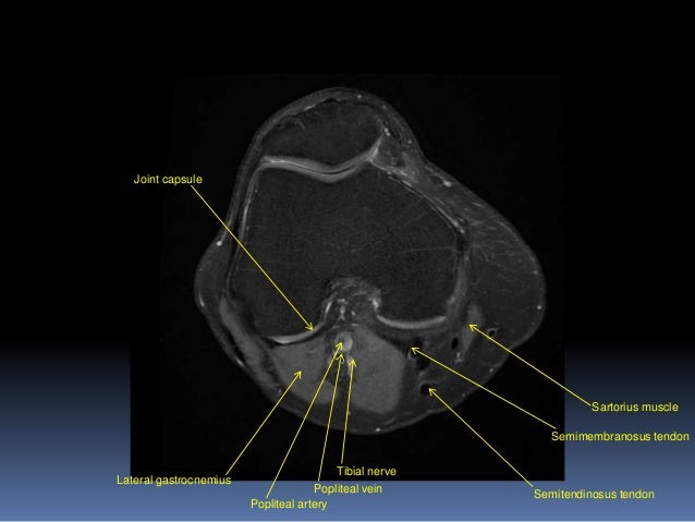
MRI Knee Anatomy from i.pinimg.com The muscles of the knee include the quadriceps, hamstrings, and the muscles of the calf. Plantaris can have variable size, but in most cases is difficult to demonstrate on routine mri studies. Articular surface of patella and femur, condyle, epicondyle and muscles (popliteus, sartorius, gastrocnemius, semimembranous with tendos.) the images obtained were exported to jpeg from dicom data stored on the pacs (picture archiving and communicating system). The normal anatomy of the knee as seen on magnetic resonance. The images may also help physicians to distinguish normal, healthy tissues from dead tissues(2). Louis, usa and the rijnland hospital in leiderdorp, the netherlands. While a detailed explanation of mri protocols and mr physics is beyond the scope of this text, fast spin echo (fse) mri is most commonly utilized for mri of the knee. In this presentation mri anatomy biceps femoris muscle.
Anatomy basic knee mri checklist.
This mri knee sagittal cross sectional anatomy tool is absolutely free to use. Louis, usa and the rijnland hospital in leiderdorp, the netherlands. In one investigation, depicted only on the proton density weighted images. T2w axial fat sat 1. Saddle joint between patella and femur; Cross sectional anatomy of the knee based on mri : Can also generate proton density images. The images may also help physicians to distinguish normal, healthy tissues from dead tissues(2). Radiology knee this app is a valuable tool for radiologists surgeons medical students doctors and nurses. Thigh muscles are responsible for allowing normal gait and proper lower extremity function (1). View of the anatomical labels. The muscles of the knee include the quadriceps, hamstrings, and the muscles of the calf. This article is based on a presentation given by david rubin and adapted for the radiology assistant by robin smithuis.